


PhD · 2027–2030
Dynamics and deformations of a biomimetic active tissue
Master 2 internship, followed by a PhD
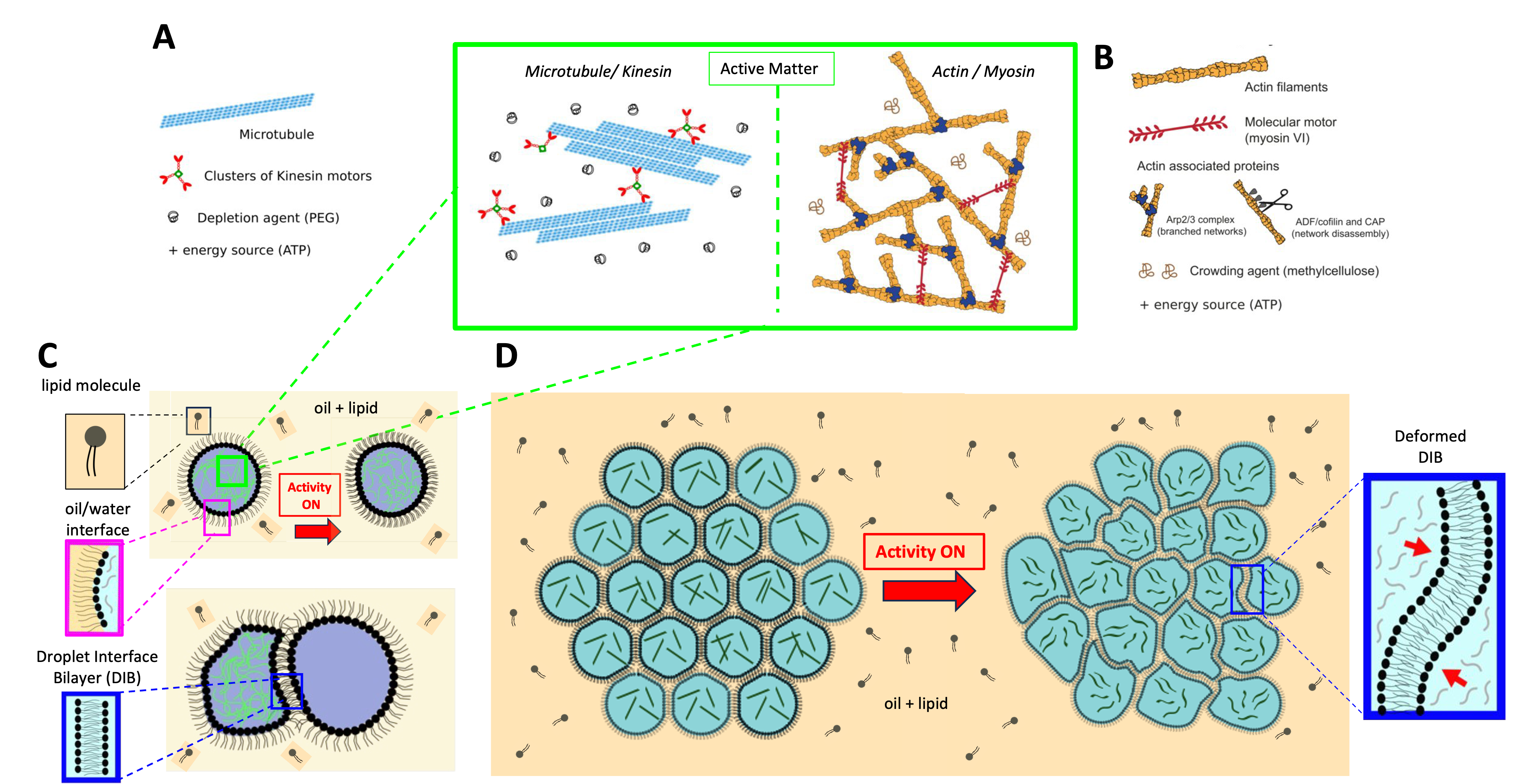
From a physics point of view, biological tissues can be studied as active matter: a collection of agents (cells) that consume ATP to perform mechanical work, and thereby deform and move. This work originates in the cytoskeleton, whose filaments are set in motion by molecular motors (actin/myosin, microtubules/kinesin). The forces generated at the cell membrane are transmitted from cell to cell through adhesion complexes. The resulting deformations, such as the folding and stretching of developing tissues, determine shape and function during morphogenesis. How cells collectively achieve this is a major question at the intersection of physics and developmental biology.
To address this challenge, biomimetic approaches have encapsulated active cytoskeletal filaments in droplets or vesicles, showing that the organization of the filament network depends on confinement and activity. These remain isolated artificial cells, without contact or adhesion. To our knowledge, no active and adhesive artificial tissue has been built.
(A) Graphical abstract of the project. (B) Fluorescence image (tubulin channel) of a circular 2D network encapsulating the microtubule–kinesin mixture. The network remains stable for several hours. (C) Fluorescence image (lipid channel) showing a T1 event (total duration: 1 h). A movie is available here.
The objective of the proposed thesis is to fabricate biomimetic active tissues, made of deformable droplets encapsulating microtubule bundles with controllable activity and adhesive between them. The tissue is a network of Droplet Interface Bilayers (DIBs): droplets immersed in an oil and lipid bath spontaneously form an adhesive lipid bilayer where they touch, the analogue of a cell–cell junction. Activity is triggered by UV uncaging of caged ATP, so that the tissue becomes active at once, only once it is fully assembled. With this system the goal is to measure and model how the activity, which depends notably on the concentrations of microtubules, kinesin motors and ATP, induces deformations of the droplets and their collective movement. The application of spatial activity gradients, using structured UV illumination, and their consequences at a larger scale will also be considered. The work is experimental; the modelling will be carried out with our theoretical collaborators.
Master 2 internship
The thesis will be preceded by an M2 internship devoted to the elementary units of the system, before any assembly. The intern will encapsulate the microtubule–kinesin network in single droplets and quantify, by fluorescence microscopy and particle image velocimetry, how the internal active flows deform the droplet interface. Bringing two droplets into contact then forms a single DIB, the analogue of a cell–cell junction, whose curvature fluctuations will be followed under activity in one or both droplets. These measurements provide the reference against which the behaviour of the full tissue will be read.
Profile sought
M2 student in biophysics or soft matter physics, with a marked taste for experimental work and for bottom-up approaches. Prior exposure to optical microscopy, image analysis, microfluidics or physico-chemistry is appreciated but not required.
Practical details
Start January–March 2027 (internship), followed by the PhD. Supervision Elie Wandersman (Prof. SU) and Jean-Christophe Galas (DR CNRS), Laboratoire Jean Perrin Apply — send a CV and a short motivation letter toelie.wandersman@sorbonne-universite.fr Copy jean-christophe.galas@sorbonne-universite.fr Copy
//